
Bunion Surgery With Morton’s Toe Shortening Explained
Bunions and Morton’s toes are common podiatric conditions – sometimes, they can be found on the same foot. If you’re struggling with both of these
At Luxe Foot Surgery, we provide the best possible service for Heel Spur Surgery in Miami. Our facilities prioritize hygiene, comfort, and state-of-the-art technology, which allows for minimally invasive procedures to reduce pain and recovery time. Our specialists are highly trained to provide ethical and professional treatment to our patients. Additionally, we offer internal payment plans and free consultations with specialists.
AVERAGE COST
$5000 - 7000
PROCEDURE TIME
30 – 40 Min
BACK TO WORK
2 - 3 week
FULL RECOVERY
3 - 6 months
Book Your Free Consultation
The heel spur is a condition of the foot, which consists of the appearance of a small bony protrusion or calcium deposit in the heel or calcaneus bone. This growth is abnormal and may take the form of a protracted beak or spearhead. It appears in the area of the heel bone, which is on the sole of the foot. The spur can extend up to half an inch.
When the calcaneal spur appears, the tissues that surround it begin to dig into it. The pressure of the body’s weight on these tissues when walking generates inflammation that can cause a lot of pain. The sensation is that of stepping on a nail every time the foot rests. The condition improves with conservative methods or with heel spur surgery.
The calcaneus is the heel bone, the large bone at the back of the foot. Here is the subtalar joint, one of the most important for locomotion, since the proper functioning of the foot depends on it. A large part of the body weight also falls on the heel bone or calcaneus when standing or moving. When the calcaneus grows abnormally and extends forward in the shape of a beak or tip, we are dealing with a calcaneal spur.
If conservative therapies, after 9 or 12 months, do not cause results, doctors proceed to perform calcaneal spur surgery. This surgery is performed after a detailed imaging study. In this way it is located exactly where the spur is, as well as what size and what shape it is. Imaging studies allow us to see which area of the tendon is affected. The heel spur operation can be done through endoscopic surgery, which requires minimal access, or through open surgery, for which a larger incision must be made. In any case, the goal of heel spur surgery is:
Heel spur surgery in Miami can cost between $4,000 and $10,000 . The money that patients have to pay out of pocket will also depend on the percentage that their health insurance can cover. The total cost of heel spur surgery varies depending on a few factors such as:
At our Luxe Foot Surgery center we maintain competitive prices compared to those of other clinics or other counties and states in the USA. In addition, we have internal payment plans, to facilitate financing for patients, so that they do not have to do without surgery for financial reasons. If you need a quote for calcaneal spur surgery, you can schedule a free appointment to start a study of your case. We’re here to serve you!
The symptomatology of the calcaneal spur is a bit irregular. There are people who have heel spurs and never feel pain. They only find out about the condition when for some other reason they take an x-ray image of the foot. For other people, heel spurs are often quite painful, especially while they are forming. When this deformity is in a critical state, it can severely limit the mobility of patients. The pain is not due to the spur itself, but to the tissues around it. Among the most common symptoms of heel spurs are:
The results of heel spur surgery improve the quality of life of patients. After the procedure, symptoms should disappear and patients will be able to regain normal foot functions. Here’s a look at some of the benefits patients get after the recovery period:
Calcaneal spurs, also known as heel spurs, are a common condition affecting many people, particularly those over 40 years of age. The condition is characterized by the formation of a bony outgrowth or protrusion in the heel bone, which can cause intense pain and discomfort. In this article, we will explore the various causes of calcaneal spurs in detail and discuss how to prevent and manage this condition.
Plantar fasciitis is a common cause of calcaneal spurs, affecting up to 80% of individuals who develop this condition. Plantar fasciitis occurs when the plantar fascia, a thick band of tissue that runs along the bottom of the foot, becomes inflamed and irritated. The condition is often caused by overuse or excessive stress on the foot, such as running, walking long distances, or standing for prolonged periods. Over time, this stress can cause micro-tears in the fascia, leading to the formation of a calcaneal spur.
The type of foot anatomy and gait disorders can also contribute to the development of calcaneal spurs. Individuals with high arches or flat feet are more prone to developing this condition, as are those with a misaligned spine or pelvic tilt. Congenital deformities of the feet, such as pes planus (flat feet) or pes cavus (high arches), can also cause imbalances in gait, leading to increased stress on the plantar fascia and the heel bone junction.
Wearing unsuitable footwear, such as flat or unsupported shoes, is another common cause of calcaneal spurs. Shoes that lack proper arch support or cushioning can cause pressure to be unevenly distributed on the foot, leading to increased stress on the heel. Narrow shoes can also contribute to an altered gait and increased pressure on the heel.
Obesity and excessive weight are two of the main risk factors for developing calcaneal spurs. Excess weight places greater stress on the plantar fascia and the heel bone junction, leading to the formation of calcium deposits over time. Individuals who are very tall, even if not overweight, may also be at increased risk of developing this condition due to the greater load placed on their feet.
Age is another risk factor for calcaneal spurs, with statistical studies showing that 11% of the US adult population has developed this condition. Calcaneal spurs have a higher incidence in active adults around 40 years of age. Adults between 60 and 70 years of age may also develop calcaneal spurs, often as a result of arthritic processes.
High-performance sports or running on hard surfaces can also contribute to the development of calcaneal spurs. These activities place prolonged stress on the foot, which can cause micro-tears in the plantar fascia and lead to the formation of calcium deposits. Many high-performance athletes may suffer from this condition, to the point of requiring surgical intervention.
The most common types of calcaneal spur surgery are the ones we will look at below. It is up to the specialist to determine which of the procedures is most appropriate for each patient. Sometimes patients can receive two of these surgical procedures during the operation. It all depends on the conditions and needs of each case.
This procedure is the most performed when the plantar spur is accompanied by plantar fasciitis. Heel spur surgery with partial or total release of the plantar fascia consists of cutting part of the tendon tissue that is attached to the heel bone. This produces some separation between the plantar fascia and the bone, which translates into a relief of tension and inflammation of the tendon.
This is the type of heel spur surgery that is usually performed when the heel spur is quite large and causes a lot of pain. It consists of completely removing the bony protuberance or spur of the heel bone. Then the bone is smoothed, so that it does not create new discomfort. It can be done by endoscopic technique or by open surgery. In many cases, heel spur removal is performed after plantar fascia release surgery, allowing better access to the heel spur.
This type of calcaneal spur surgery consists of performing a surgical lengthening of the calf muscles. When the gastrocnemius muscle (popularly known as the “calf muscle”) is tense, it puts more pressure on the plantar fascia. Through this surgery one of the calf muscles is lengthened, with the aim of increasing movement and flexion of the ankle. Depending on the conditions, the surgery can be performed through an open (traditional) incision or with minimally invasive technology. It has a low complication rate. It is generally applied to patients who have great difficulty in flexing the foot.
After heel spur surgery, patients must go through a recovery period that can be divided into different stages. The success of recovery depends to a large extent on patients fully complying with the care recommended for each stage. As the days and months go by, the foot will gradually recover its strength and functions, until it finally returns to normality. The length of time it takes to recover varies depending on the type of heel spur surgery performed. Let’s examine the necessary care in each stage of recovery, after calcaneal spur surgery.
After the operation, the foot will be completely bandaged, and you may have a cast, boot or other immobilization accessory. When leaving the hospital, patients must rest completely for the rest of the day. The most advisable thing is not to bathe that same day. The optimal thing is to remain lying down with the foot high, on pillows. It is recommended that you leave the hospital with a companion and also stay accompanied at home.
It is normal to feel pain in the foot during the first days after surgery. To avoid pain and inflammation, the surgeon will prescribe a plan of analgesics and anti-inflammatories. Applying ice packs is a good way to prevent swelling and bruising. Ice should not be on the wound area for more than 15 minutes, but it can be applied several times a day. It should also not be applied directly to the skin.
The patient should not stand on the foot during the first 2 weeks of recovery. If you need to get up, you must keep your foot off the floor. To achieve this you can use crutches, a wheelchair, or other accessories. When you go to bathe, you should keep the bandage dry and clean.
After the first two weeks, even a little earlier, at 10 days, the patient will have an appointment with the surgeon to have the stitches removed. You may have an x-ray or CT scan at this appointment. By this time the incision should be completely healed. The patient can begin to support the foot to walk. Physiotherapy exercises will help tone muscles. Those who have office work or some other type of job that requires sitting will be able to join at this time, although they will still have to use boots or crutches to walk. On the other hand, people who work standing up must wait until the fourth week to start work.
From the sixth week to three months after surgery, the patient’s full recovery is expected to occur. With physiotherapy exercises and walking practice, the foot will be stronger every day. By the third month, it is expected that the pain will disappear and that there will be a complete resolution of the symptoms of the calcaneal spur. It is recommended to continue using appropriate footwear, to avoid the reappearance of the spur.
Heel spur surgery is the solution for those patients who have already tried conservative methods and have not obtained the expected results, or have not perceived any improvement. To perform calcaneal spur surgery, the condition must present the most severe symptoms, which prevent a normal life. In general, these symptoms are based on feeling a lot of pain when supporting the foot, so that the patient is forced to modify his posture to avoid that the weight falls on the heel. In these cases, a heel spur surgery avoids the consequences that adopting a bad posture can have on all the joints.
The preoperative evaluation of the heel spur goes through two main phases: one is the medical examination and the other is the tests. Before calcaneal spur surgery, it is important to perform a medical examination in order to diagnose the condition. First the patients must describe the symptoms, and then the specialist will look for some signs, such as:
Clinical studies are completed with imaging tests. A lateral calcaneal X-ray will reveal the protrusion called a heel spur, what shape it is, and how long it is. X-rays provide clear images of the bones, which can rule out pain from fractures or arthritis. MRI tests and ultrasound are usually not used for heel spurs. Also before surgery, blood samples are taken to analyze and rule out diseases such as rheumatoid arthritis, ankylosing spondylitis or gout.
Before the operation, patients meet with the surgeon for a review of tests and studies. Here it is determined which type of heel spur surgery is most appropriate. The doctor will give specific instructions to the patient regarding what to stop doing before and after the operation. General recommendations:
Heel spur surgery is not without risk, just like any other surgery. However, it should be noted that the percentage of people who present problems after this type of surgery is very low. However, it is advisable for patients to be aware of these possible risks before undergoing the procedure. Next, we will see some of the most common problems that heel spur surgery can cause.
Although most patients who undergo heel spur surgery gradually become pain-free, until the pain disappears for good, there is a risk of continued pain beyond the first week of recovery. This chronic pain can be temporary and disappear after a while, or stay permanently.
When performing heel spur surgery, there is some risk of injury to the foot nerve or nerve branches. This can cause a numbness or tingling sensation around the operated area. Sometimes the nerve damage is temporary, so patients will regain feeling after a while. However, in some cases the damage can be permanent.
This is one of the most unwanted risks of all surgery. Clots can form in the area where the surgery was performed, or form in another place as a result of the period of inactivity. It is likely that the surgeon will prescribe the patient a plan with heparin, to prevent clot formation, especially in patients with a genetic history of vascular risks. Clots should be avoided at all costs, as if they travel through the bloodstream they can cause a pulmonary or cerebral embolism.
Although heel spur surgery is performed in an antiseptic environment, making a surgical incision always carries the risk of infection. This is because the protective barrier against the entry of pathogens that is the skin is broken. The best way to avoid an infection is to follow the instructions that the doctor will give about the care and hygiene that must be taken with the wound during the recovery period. A plan of antibiotics to take after heel spur surgery will probably also be indicated.
When heel spur surgery involves the release of the plantar fascia (one of the procedures described above) there is a risk that the structure of the foot will be modified. The foot can undergo a permanent change in its morphology. The plantar fascia is the tendon or ligament that supports the foot when the person is standing, walking or running. It is also the one that naturally forms the plantar arch. With surgery, the plantar fascia can become loose, putting you at risk of losing part of the support mechanism. Functions such as cushioning and releasing loads are weakened. This can cause problems like:
One of the risks of percutaneous calcaneal spur surgeries is heel fracture. Although the percentage of risk of this happening is really very low. Patients who return home with a fractured heel may experience a popping sensation when they try to walk on their foot, pain, and functional impotence. In these cases, another surgery will be performed to remedy the fracture by using screws. The goal is for the bone to perform osteosynthesis.
Heel spur surgery is usually done on an outpatient basis, so most patients can go home the same day. The patient must be prepared to receive some type of anesthesia that can be: general, regional or local. The recovery period will be shorter for endoscopic or minimal access surgeries than for open ones. When you leave the operating room, your foot may be bandaged or have a splint, cast, or boot.
When the bone structure called calcaneal spur is removed, the symptoms that afflicted patients usually disappear. While the recovery process takes place, those who have received heel spur surgery will gradually recover their foot functions to normal.
Yes, there are several non-surgical treatments for heel spurs. Before performing heel spur surgery, one or more of the conservative methods of treating this ailment are tried. Actually, calcaneal spur surgery is the last resort that specialists propose to their patients. Although this type of non-surgical treatment does not eliminate the heel spur, it can greatly improve its symptoms. It is estimated that 90% of patients see improvement in this condition after 10 months practicing non-surgical treatments. Let’s now see some of the most used conservative or non-surgical methods to improve the condition of heel spurs.
One of the treatments that improve the condition of those who suffer from heel spurs is to wear thick-soled shoes with good cushioning. If the shoe has good padding and a pad is added that covers the heel area, the tension on the fascia is reduced when walking, and as a consequence, microtraumatisms are reduced. This can greatly relieve pain from walking or standing. As a recommendation, patients should stop wearing worn-out shoes that do not have support. Athletic shoes should also be replaced before their soles wear out.
Some accessories can be purchased on the market to provide greater support and cushion the foot when walking. Soft silicone pads protect the heel area while raising and cushioning it. You can also use an orthopedic insole of those that are made to measure, according to the conditions and needs of the patient. Another recommended device is night splints. Most people sleep with their feet pointing down, a position that relaxes the plantar fascia. With the splint, the foot assumes a position that keeps the fascia stretched throughout the night. It can be a little hard to get used to, but the splint reduces the pain caused by plantar fasciitis.
Another option to improve the symptoms of heel spurs is to follow a physical therapy exercise plan. The exercises will be performed with the help of a physical therapist and will focus on stretching the calf muscles and plantar fascia. It may also include ice therapy and massage to decrease inflammation of the fascia.
When patients start to feel pain, it can be relieved by taking non-steroidal anti-inflammatory drugs (NSAIDs). The most common are ibuprofen and naproxen. The use of these medicines should not exceed a month, since both can cause side effects such as ulcers, bleeding in the stomach or intestines, constipation, excess gas, thirst, headache, etc.
When heel spurs are accompanied by plantar fasciitis, your doctor may recommend immobilizing your foot for a while. A cast will be placed that will keep the foot immobile, with the aim of deflating the plantar fascia. This can also be done with special boots, but a cast is more effective. The cast keeps the foot 100% immobile, while the patient can remove and put on the boots himself, which reduces the effectiveness of the treatment.
Cortisone is a powerful anti-inflammatory steroid that is injected directly into the plantar fascia. It has the effect of reducing inflammation and pain. Although it is a very effective procedure, doctors tend to use it very sparingly, because the use of cortisone can weaken the plantar fascia to the point of tearing the plantar fascia. This is very dangerous, because it changes the structure of the foot and can cause chronic pain.
Another injection that is much less risky than cortisone is platelet-rich plasma (PRP). This substance is obtained from the blood of patients, and its objective is to promote the regeneration of the plantar fascia. The downside is that it is a more expensive treatment and is still under investigation.
Extracorporeal Shock Wave Treatment (ESWT) is another non-surgical method for treating heel spurs. It consists of stimulating the healing process of damaged tissues in the plantar fascia through the use of high energy shock waves. It is a fairly safe and non-invasive procedure, which is still being investigated.
It is a minimally invasive treatment. It is used as a resource before performing surgery in those patients who have persistent plantar fasciitis. The procedure is performed using ultrasound imaging as a guide to insert a probe into the injured plantar fascia. The probe tip emits vibrations that break up and remove damaged tissue.
Our Luxe Foot Surgery patients often ask a few questions about heel spur surgery. Next, we will answer some of the most frequent questions. If you have a question that is not included in this article, we recommend that you contact our center by phone or through social networks. Our advisors will gladly assist you. If you need to evaluate your case with a specialist, remember that you can also schedule a free appointment .
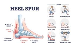
They are not the same. The plantar fascia is the ligament that connects the heel to the forefoot, and plantar fasciitis is inflammation of the plantar fascia. On the other hand, the heel spur is an abnormal protrusion of the heel bone that appears in the area of the sole of the foot. These are two different conditions. Many patients tend to confuse them because, in a high percentage of cases, they appear together, because plantar fasciitis can be one of the causes of heel spurs and vice versa. However, having plantar fasciitis does not mean that there will be a spur, and that there is a spur does not mean that fasciitis has to appear.
Those who suffer from heel spurs can perform several of the conservative treatments at home, without the need to go to any hospital. Among the most common home methods for heel spurs we have the following:
Most people with heel spurs improve their condition with conservative treatment. But when heel spurs are not treated in time, the condition can become chronic and worsen your symptoms. This implies feeling more pain and experiencing difficulties to support the foot when walking.
Benjamin Franklin was quite right when he said “An ounce of prevention is worth a pound of cure”. This phrase can be applied to heel spurs as well. To prevent is to change those factors that contribute to the formation of the calcaneal spur. Some prevention actions are in our hands:
Not all patients can undergo heel spur surgery, as there are contraindications for some cases. Although there are few contraindications, not complying with them could expose the health of patients. However, it is up to the specialist to carry out a detailed study of each case, to measure the seriousness or not of their situation. Patients who:
When you go to an appointment with the specialist for calcaneal spurs, you can ask him some questions that allow you to better understand this condition. Some questions you can ask are:
We also recommend that the person notify their doctor of any type of condition they have, as this will be taken into account to avoid risks in calcaneal spur surgery. It is of the utmost importance to state whether the potential patient has had a family history of deep vein thrombosis (DVT).
The pain felt in heel spur surgery is minimal, since the foot will be anesthetized. However, you may feel pain when the anesthesia wears off and for the first few days. But that pain will disappear as the foot recovers. Resting, keeping your foot elevated, placing ice packs around the incision area, and even taking pain relievers can greatly relieve pain. Another time when the patient may feel pain again is when they begin to support the foot to walk after surgery.
During the recovery period, patients should observe the evolution of the wound daily. And they should contact their doctor if they experience any of the following symptoms:
Heel spur surgery has a high success rate, along with a low risk rate. Only a low percentage of those operated on have calcaneal spur problems again. For most patients, with surgery the spur disappears completely, along with the pain and swelling when walking. Although heel spur surgery has been effective, patients must try not to return to the same habits, in order to avoid the appearance of new heel spurs.
This will depend on the type of heel spur surgery that has been performed and how quickly patients recover. The fastest time to start walking is 10-15 days after the stitches are removed from the scar. There will be patients who must wait a little longer, and in those cases it will be the specialist who will indicate the precise moment. The maximum time to start walking can be 3 months.
Before driving again, the patient should ask his surgeon if he thinks he is ready for that activity. Generally, if the heel spur surgery is on the foot that is not used for driving (in automatic cars), the patient may be able to do it again after the first week of recovery. However, it is best to wait until the surgeon has removed the stitches before driving again. We also recommend asking the car insurance what protocols they have for recently operated drivers in the event of an accident.
No. Once a heel spur appears, it does not go away on its own. Not all heel spurs are painful or cause any symptoms. Even, as we have said, the majority of patients who present symptoms experience an improvement with conservative methods, so they will not need surgery and will be able to live with the calcaneal spur. The group of patients who do not find improvement with non-surgical methods should undergo surgery. The heel spur operation is the only way that the protuberance does disappear, since it is removed during the procedure and the bone is left smooth.
After the first 2 weeks of recovery after heel spur surgery, most patients can begin to walk. At that time, it is likely that there will be pain in the foot muscles and that the person will not feel very strong to hold on. This is normal, because the muscles and tendons will be weak after so many days of rest, and also because of the surgical procedure itself. Most likely, the doctor will indicate physical therapy exercises to strengthen the foot. Among these exercises we have:
A greater accumulation of calcium salts and unmetabolized uric acid make the body more prone to calcaneal spurs. That is why some dietary modifications can prevent the mole from growing further.
After heel spur surgery, it is normal to have swelling. In fact, the swelling in the foot, especially in the area of the incision, can last several weeks. The inflammation will disappear as the foot recovers. However, patients should be observant, as exaggerated and persistent swelling, including redness and pain, is not normal. In such cases, you should see a surgeon as soon as possible.
Bunions and Morton’s toes are common podiatric conditions – sometimes, they can be found on the same foot. If you’re struggling with both of these
A bunion is an unpleasant foot deformity that can only be permanently corrected with surgery – but what can you expect after that procedure? If
A bunion is one of the most painful foot deformities – it often affects everyday activities and makes it difficult to walk or run. Luckily,
The bunion is a complex deformity – it can get progressively more painful over time, and at some point, you will realize that you need
Many people struggle with a bunion on the little toe – this condition can cause unpleasant symptoms and interfere with your everyday life. Before you
What can you do when your bunion has been causing discomfort and pain throughout the entire day? As soon as you take off your tight
Are you exhausted from dealing with the pain and discomfort caused by bunions? Seeking an adequate pair of shoes might be trickier than you imagine.
If you’re having minimally invasive bunion surgery soon, you must be wondering what the recovery process will look like. The four-week mark is an important
How to tell if the shape of your foot is normal or if you have a bunion? It can be easy to confuse a slightly
Monday – Thursday: 9AM – 7PM Friday – Saturday: 9AM – 7PM Sunday: Closed
Copyright © 2022 All Rights Reserved